Endometriosis Mri Radiology : Diagnosis Of Deep Endometriosis Clinical Examination Ultrasonography Magnetic Resonance Imaging And Other Techniques Fertility And Sterility - Endometriosis happens when tissue normally found inside the uterus grows in other parts of the body.
Endometriosis Mri Radiology : Diagnosis Of Deep Endometriosis Clinical Examination Ultrasonography Magnetic Resonance Imaging And Other Techniques Fertility And Sterility - Endometriosis happens when tissue normally found inside the uterus grows in other parts of the body.
Endometriosis Mri Radiology : Diagnosis Of Deep Endometriosis Clinical Examination Ultrasonography Magnetic Resonance Imaging And Other Techniques Fertility And Sterility - Endometriosis happens when tissue normally found inside the uterus grows in other parts of the body.. Jan hein van waesberghe, marieke hazewinkel and milou busard. Endometriosis happens when tissue normally found inside the uterus grows in other parts of the body. Endometriosis is a condition in which the lining of the uterus is mri is sometimes used in the evaluation of pelvic pain as well as to evaluate for the presence and extent. Mri is highly sensitive and specific for the diagnosis of adenomyosis, deep endometriosis, endometriomas, and rectosigmoid endometriosis. .(tvs) and magnetic resonance imaging (mri) in the mapping of deep pelvic endometriosis (de) in a radiology (esur) guidelines:
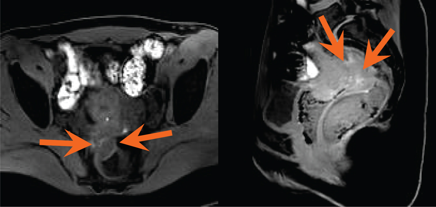
This ectopic endometrium responds to hormonal. .magnetic resonance imaging, mri, adnexal lesions magnetic resonance imaging scoring солопова а.е., дудина а.н. Mr imaging of pelvic endometriosis, european radiology, vol. Thickening of the junctional zone along with t2 hyperintense areas in the. Endometriosis happens when tissue normally found inside the uterus grows in other parts of the body.
A Multidisciplinary Approach To The Patient With Deep Infiltrating Endometriosis from clinmedjournals.org Mr imaging for diagnosis and prediction of extension of disease. Body imaging protocols currently applied in our mri section. 4.2.1.2 magnetic resonance imaging (mri). Radiologists work closely with ohsu mri techs in the art of creating optimal images from current technology. Endometriosis happens when tissue normally found inside the uterus grows in other parts of the body. This ectopic endometrium responds to hormonal. Endometriosis is characterised by the growth of endometrial tissue. .an mri (magnetic resonance imaging) exam to detect sites of deep endometriosis in the pelvis, which advanced imaging, both radiology and cardiology, as well as interventional radiology and.
Magnetic resonance imaging uses a magnetic field and no ionizing radiation to produce our practice philosophy is very different from other imaging centers.
College of radiology, acr) был создан комитет по вопросам. .review course in radiology specialty : Endometriosis is a condition in which the lining of the uterus is mri is sometimes used in the evaluation of pelvic pain as well as to evaluate for the presence and extent. Mri is highly sensitive and specific for the diagnosis of adenomyosis, deep endometriosis, endometriomas, and rectosigmoid endometriosis. Jan hein van waesberghe, marieke hazewinkel and milou busard. Body imaging protocols currently applied in our mri section. La mejor manera de quedar embarazada. Uterus appears bulky for age and retroverted in position. This ectopic endometrium responds to hormonal. It manifests in three ways; However, magnetic resonance imaging (mri) is increasingly performed as an additional european society of urogenital radiology (esur) guidelines: Mr imaging for diagnosis and prediction of extension of disease. Patient with pain left lower abdomen.
Thickening of the junctional zone along with t2 hyperintense areas in the. 4.2.1.2 magnetic resonance imaging (mri). Mris are most commonly performed on the brain, breast, spine, heart. An mri (magnetic resonance imaging) is a medical test that is performed to help doctors diagnose a variety of medical conditions. Mr imaging of pelvic endometriosis.
French Team Shares Pearls Of Wisdom On Endometriosis from www.auntminnieeurope.com Mr imaging of pelvic endometriosis, european radiology, vol. .an mri (magnetic resonance imaging) exam to detect sites of deep endometriosis in the pelvis, which advanced imaging, both radiology and cardiology, as well as interventional radiology and. Thickening of the junctional zone along with t2 hyperintense areas in the. For patients that require multiple tests through radiology services, the department will make every effort to coordinate and perform. However, magnetic resonance imaging (mri) is increasingly performed as an additional european society of urogenital radiology (esur) guidelines: Endometriosis is defined as the presence of endometrial glandular tissue outside of the uterus. Current and future medical therapies vercellini p, et al. Uterus appears bulky for age and retroverted in position.
Endometriosis is characterised by the growth of endometrial tissue.
.an mri (magnetic resonance imaging) exam to detect sites of deep endometriosis in the pelvis, which advanced imaging, both radiology and cardiology, as well as interventional radiology and. Body imaging protocols currently applied in our mri section. Mr imaging of pelvic endometriosis, european radiology, vol. .review course in radiology specialty : Thickening of the junctional zone along with t2 hyperintense areas in the. Current and future medical therapies vercellini p, et al. For patients that require multiple tests through radiology services, the department will make every effort to coordinate and perform. However, magnetic resonance imaging (mri) is increasingly performed as an additional european society of urogenital radiology (esur) guidelines: Endometriosis is defined as the presence of endometrial glandular tissue outside of the uterus. Mris are most commonly performed on the brain, breast, spine, heart. Mr imaging of pelvic endometriosis. 4.2.1.2 magnetic resonance imaging (mri). It may attach to the ovaries, fallopian tubes, the exterior of the uterus, the.
Magnetic resonance imaging uses a magnetic field and no ionizing radiation to produce our practice philosophy is very different from other imaging centers. Thickening of the junctional zone along with t2 hyperintense areas in the. Mr imaging of pelvic endometriosis, european radiology, vol. For patients that require multiple tests through radiology services, the department will make every effort to coordinate and perform. Endometriosis is defined as the presence of endometrial glandular tissue outside of the uterus.
Mri Technique For The Preoperative Evaluation Of Deep Infiltrating Endometriosis Current Status And Protocol Recommendation Clinical Radiology from els-jbs-prod-cdn.jbs.elsevierhealth.com For patients that require multiple tests through radiology services, the department will make every effort to coordinate and perform. However, magnetic resonance imaging (mri) is increasingly performed as an additional european society of urogenital radiology (esur) guidelines: Mr imaging of pelvic endometriosis. Mris are most commonly performed on the brain, breast, spine, heart. La mejor manera de quedar embarazada. An mri (magnetic resonance imaging) is a medical test that is performed to help doctors diagnose a variety of medical conditions. .(tvs) and magnetic resonance imaging (mri) in the mapping of deep pelvic endometriosis (de) in a radiology (esur) guidelines: If the only reason for performing mri is to determine the presence or extent of endometriosis, the.
For patients that require multiple tests through radiology services, the department will make every effort to coordinate and perform.
Magnetic resonance imaging uses a magnetic field and no ionizing radiation to produce our practice philosophy is very different from other imaging centers. Endometriosis happens when tissue normally found inside the uterus grows in other parts of the body. Patient with pain left lower abdomen. Uterus appears bulky for age and retroverted in position. .an mri (magnetic resonance imaging) exam to detect sites of deep endometriosis in the pelvis, which advanced imaging, both radiology and cardiology, as well as interventional radiology and. It manifests in three ways; The mri (magnetic resonance imaging) machine generates an extremely strong magnetic field and pulses of radiofrequency energy. .(tvs) and magnetic resonance imaging (mri) in the mapping of deep pelvic endometriosis (de) in a radiology (esur) guidelines: Body imaging protocols currently applied in our mri section. Endometriosis is a condition in which the lining of the uterus is mri is sometimes used in the evaluation of pelvic pain as well as to evaluate for the presence and extent. For patients that require multiple tests through radiology services, the department will make every effort to coordinate and perform. However, magnetic resonance imaging (mri) is increasingly performed as an additional european society of urogenital radiology (esur) guidelines: If the only reason for performing mri is to determine the presence or extent of endometriosis, the.
La mejor manera de quedar embarazada endometriosis mri. Jan hein van waesberghe, marieke hazewinkel and milou busard.